
















©The Author(s) 2017.
World J Gastroenterol. Dec 28, 2017; 23(48): 8671-8678
Published online Dec 28, 2017. doi: 10.3748/wjg.v23.i48.8671
Published online Dec 28, 2017. doi: 10.3748/wjg.v23.i48.8671
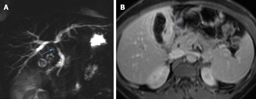
Figure 1 Preoperative magnetic resonance cholangiopancreatography from case 1 demonstrates stenosis of the common bile duct and biliary confluence (arrow, A) and retrograde biliary dilatation.
The transverse section demonstrates diffuse asymmetrical gallbladder wall thickening (arrowhead, B) and contiguous hilar mass.
- Citation: Nacif LS, Hessheimer AJ, Rodríguez Gómez S, Montironi C, Fondevila C. Infiltrative xanthogranulomatous cholecystitis mimicking aggressive gallbladder carcinoma: A diagnostic and therapeutic dilemma. World J Gastroenterol 2017; 23(48): 8671-8678
- URL: https://www.wjgnet.com/1007-9327/full/v23/i48/8671.htm
- DOI: https://dx.doi.org/10.3748/wjg.v23.i48.8671