
















©The Author(s) 2017.
World J Gastroenterol. Dec 14, 2017; 23(46): 8152-8168
Published online Dec 14, 2017. doi: 10.3748/wjg.v23.i46.8152
Published online Dec 14, 2017. doi: 10.3748/wjg.v23.i46.8152
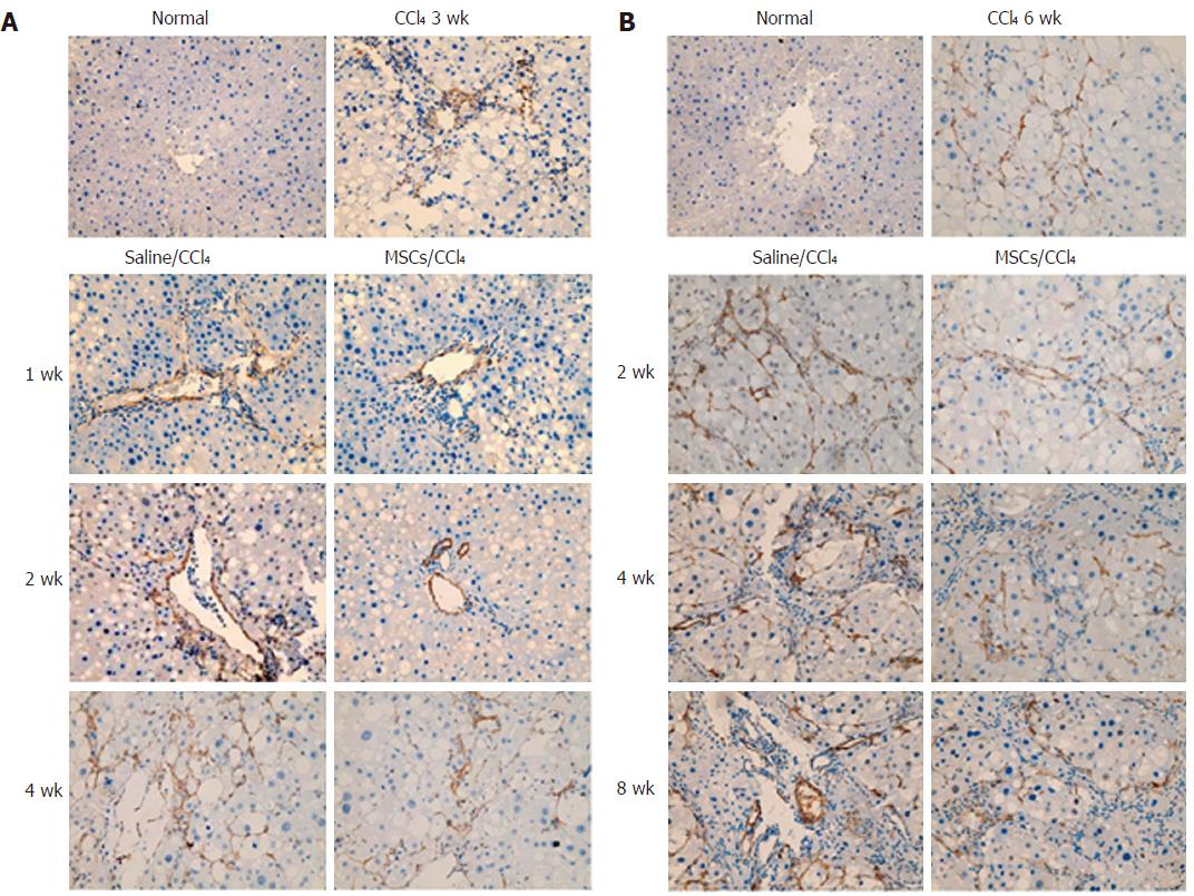
Figure 5 Immunohistochemcial staining for α-smooth muscle actin in hepatic fibrosis and cirrhosis groups (× 400).
A: Immunohistochemcial staining for α-smooth muscle actin (α-SMA) in hepatic fibrosis groups; B: Immunohistochemcial staining for α-SMA in hepatic cirrhosis groups. In normal rat liver, α-SMA was occasionally detected in vascular smooth muscle cells, and the expression level was low, revealing few activated hepatic stellate cells (HSCs). After CCl4 administration, the α-SMA spread to the portal area, showing more activated HSCs. The expression of α-SMA in liver tissues increased significantly in the saline infusion group compared with the normal group. Compared with the saline infusion groups, they significantly decreased in the MSC transplantation groups.
- Citation: Zhang GZ, Sun HC, Zheng LB, Guo JB, Zhang XL. In vivo hepatic differentiation potential of human umbilical cord-derived mesenchymal stem cells: Therapeutic effect on liver fibrosis/cirrhosis. World J Gastroenterol 2017; 23(46): 8152-8168
- URL: https://www.wjgnet.com/1007-9327/full/v23/i46/8152.htm
- DOI: https://dx.doi.org/10.3748/wjg.v23.i46.8152