Copyright
©The Author(s) 2017.
World J Gastroenterol. Oct 7, 2017; 23(37): 6802-6816
Published online Oct 7, 2017. doi: 10.3748/wjg.v23.i37.6802
Published online Oct 7, 2017. doi: 10.3748/wjg.v23.i37.6802
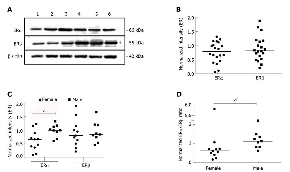
Figure 1 Expression of estrogen receptor subtypes in whole tissue lysates from normal male and female subjects.
A: Whole tissue lysates from liver tissues of normal donors were subjected to Western blotting and probed with antibodies against ERα, ERβ and β-actin. A representative blot is shown. B-D: The bands corresponding to ERα, ERβ and β-actin were quantified by densitometric analyses using ImageJ. Each symbol represents one individual. Expression of the ER subtypes was normalized to the expression of β-actin and plotted (B). Gender-based ER subtype expression was evaluated by segregating the gender and plotted (C). The ERα:ERβ expression ratio was also plotted for each gender group (D). aP < 0.05 was considered significant. ER: Estrogen receptor.
- Citation: Iyer JK, Kalra M, Kaul A, Payton ME, Kaul R. Estrogen receptor expression in chronic hepatitis C and hepatocellular carcinoma pathogenesis. World J Gastroenterol 2017; 23(37): 6802-6816
- URL: https://www.wjgnet.com/1007-9327/full/v23/i37/6802.htm
- DOI: https://dx.doi.org/10.3748/wjg.v23.i37.6802


