
















©The Author(s) 2017.
World J Gastroenterol. Mar 14, 2017; 23(10): 1735-1746
Published online Mar 14, 2017. doi: 10.3748/wjg.v23.i10.1735
Published online Mar 14, 2017. doi: 10.3748/wjg.v23.i10.1735
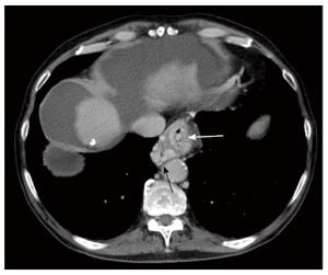
Figure 3 Axial enhanced computed tomography of the upper abdomen in portal venous phase demonstrates multiple large tubular and serpiginous esophageal (white arrow) and paraesophageal (black arrow) varices at the level of the esophageal hiatus of the diaphragm.
- Citation: Bandali MF, Mirakhur A, Lee EW, Ferris MC, Sadler DJ, Gray RR, Wong JK. Portal hypertension: Imaging of portosystemic collateral pathways and associated image-guided therapy. World J Gastroenterol 2017; 23(10): 1735-1746
- URL: https://www.wjgnet.com/1007-9327/full/v23/i10/1735.htm
- DOI: https://dx.doi.org/10.3748/wjg.v23.i10.1735