
















©The Author(s) 2016.
World J Gastroenterol. Feb 28, 2016; 22(8): 2642-2646
Published online Feb 28, 2016. doi: 10.3748/wjg.v22.i8.2642
Published online Feb 28, 2016. doi: 10.3748/wjg.v22.i8.2642
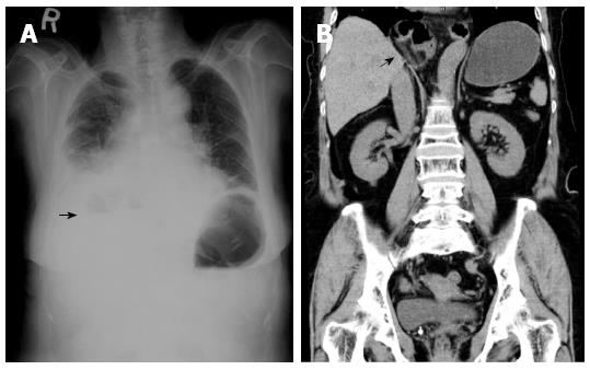
Figure 1 Plain chest radiography computed tomography of the patient.
Chest radiograph (A) revealed an intrathoracic intestinal gas bubble and an air fluid level (arrow). Thoracoabdominal computed tomography (B) confirmed an incarceration of small intestine in right thoracic cavity (arrow).
- Citation: Hsu CT, Hsiao PJ, Chiu CC, Chan JS, Lin YF, Lo YH, Hsiao CJ. Terminal ileum gangrene secondary to a type IV paraesophageal hernia. World J Gastroenterol 2016; 22(8): 2642-2646
- URL: https://www.wjgnet.com/1007-9327/full/v22/i8/2642.htm
- DOI: https://dx.doi.org/10.3748/wjg.v22.i8.2642