
















©The Author(s) 2016.
World J Gastroenterol. Dec 14, 2016; 22(46): 10131-10139
Published online Dec 14, 2016. doi: 10.3748/wjg.v22.i46.10131
Published online Dec 14, 2016. doi: 10.3748/wjg.v22.i46.10131
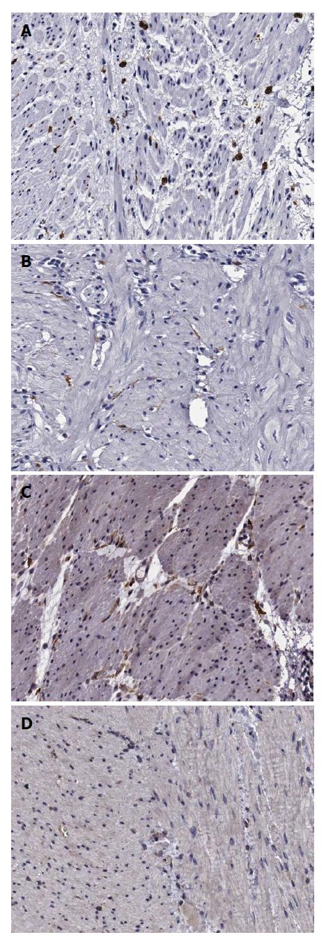
Figure 3 Immunohistochemical staining of interstitial cells of Cajal (upper panel) and platelet-derived growth factor receptor α-positive fibroblast-like cells (lower panel) in the human gastric corpus (× 200).
Cellularity of ICC is higher in the control group (A) than in the DM group (B). Cellularity of FLCs is higher in the control group (C) than in the DM group (D). DM: Diabetes mellitus; FLCs: Fibroblast-like cells.
- Citation: Park KS, Cho KB, Hwang IS, Park JH, Jang BI, Kim KO, Jeon SW, Kim ES, Park CS, Kwon JG. Characterization of smooth muscle, enteric nerve, interstitial cells of Cajal, and fibroblast-like cells in the gastric musculature of patients with diabetes mellitus. World J Gastroenterol 2016; 22(46): 10131-10139
- URL: https://www.wjgnet.com/1007-9327/full/v22/i46/10131.htm
- DOI: https://dx.doi.org/10.3748/wjg.v22.i46.10131