
















©The Author(s) 2016.
World J Gastroenterol. Jan 21, 2016; 22(3): 917-932
Published online Jan 21, 2016. doi: 10.3748/wjg.v22.i3.917
Published online Jan 21, 2016. doi: 10.3748/wjg.v22.i3.917
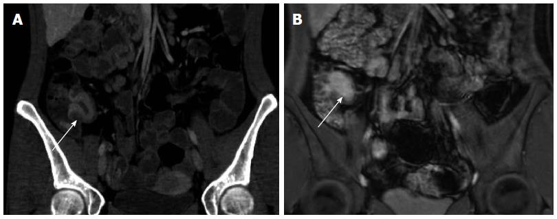
Figure 3 Active Crohn’s terminal ileitis depicted on computed tomography enterography and magnetic resonance enterography in the same patient.
Serial computed tomography enterography (A) and magnetic resonance enterography (B) studies demonstrate marked wall thickening and hyperenhancement (arrows) just proximal to the ileocecal valve consistent with active disease, as confirmed by endoscopy.
- Citation: Kilcoyne A, Kaplan JL, Gee MS. Inflammatory bowel disease imaging: Current practice and future directions. World J Gastroenterol 2016; 22(3): 917-932
- URL: https://www.wjgnet.com/1007-9327/full/v22/i3/917.htm
- DOI: https://dx.doi.org/10.3748/wjg.v22.i3.917