
















©The Author(s) 2016.
World J Gastroenterol. Jun 28, 2016; 22(24): 5598-5608
Published online Jun 28, 2016. doi: 10.3748/wjg.v22.i24.5598
Published online Jun 28, 2016. doi: 10.3748/wjg.v22.i24.5598
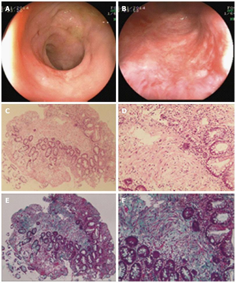
Figure 4 Histo-pathological features after colostomy in case 3.
Complete remission of confluent telangiectasia, edema and ulcer were observed 31 mo after colostomy (A and B). Diffused chronic inflammatory cells and rebuilt of mucosal integrity were seen in hematoxylin and eosin-stained slides (C: × 100; D: × 400). Massive fibrotic collagen depositions were observed in the sub-mucosa layer, with green color by Masson staining (E: × 100; F: × 400).
- Citation: Yuan ZX, Ma TH, Wang HM, Zhong QH, Yu XH, Qin QY, Wang JP, Wang L. Colostomy is a simple and effective procedure for severe chronic radiation proctitis. World J Gastroenterol 2016; 22(24): 5598-5608
- URL: https://www.wjgnet.com/1007-9327/full/v22/i24/5598.htm
- DOI: https://dx.doi.org/10.3748/wjg.v22.i24.5598