
















©The Author(s) 2016.
World J Gastroenterol. Mar 14, 2016; 22(10): 3052-3055
Published online Mar 14, 2016. doi: 10.3748/wjg.v22.i10.3052
Published online Mar 14, 2016. doi: 10.3748/wjg.v22.i10.3052
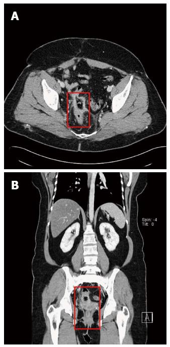
Figure 1 Computed tomography during venous phase of contrast injection.
Each red rectangle in axial (A) and coronal (B) computed tomographic images indicates wall thickening and mucosal enhancement of the rectosigmoid colon.
- Citation: Shin WY, Im CH, Choi SK, Choe YM, Kim KR. Transmural penetration of sigmoid colon and rectum by retained surgical sponge after hysterectomy. World J Gastroenterol 2016; 22(10): 3052-3055
- URL: https://www.wjgnet.com/1007-9327/full/v22/i10/3052.htm
- DOI: https://dx.doi.org/10.3748/wjg.v22.i10.3052