
















©The Author(s) 2015.
World J Gastroenterol. Feb 7, 2015; 21(5): 1684-1688
Published online Feb 7, 2015. doi: 10.3748/wjg.v21.i5.1684
Published online Feb 7, 2015. doi: 10.3748/wjg.v21.i5.1684
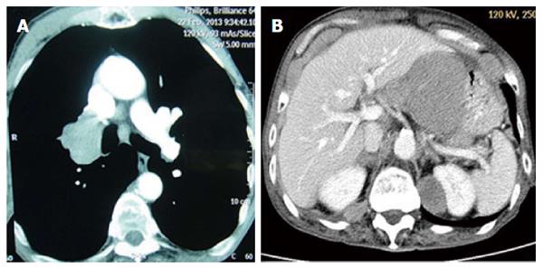
Figure 1 Computed tomography scan of the chest cut disclosed a large space-occupying lesion (5.
0 cm × 4.0 cm) at the right hilum (A), computed tomography scan of the abdomen cut showed abnormal thickening in the stomach wall, as well as lymph node tumefaction and integration (B).
- Citation: Gao S, Hu XD, Wang SZ, Liu N, Zhao W, Yu QX, Hou WH, Yuan SH. Gastric metastasis from small cell lung cancer: A case report. World J Gastroenterol 2015; 21(5): 1684-1688
- URL: https://www.wjgnet.com/1007-9327/full/v21/i5/1684.htm
- DOI: https://dx.doi.org/10.3748/wjg.v21.i5.1684