
















©The Author(s) 2015.
World J Gastroenterol. Dec 7, 2015; 21(45): 12767-12777
Published online Dec 7, 2015. doi: 10.3748/wjg.v21.i45.12767
Published online Dec 7, 2015. doi: 10.3748/wjg.v21.i45.12767
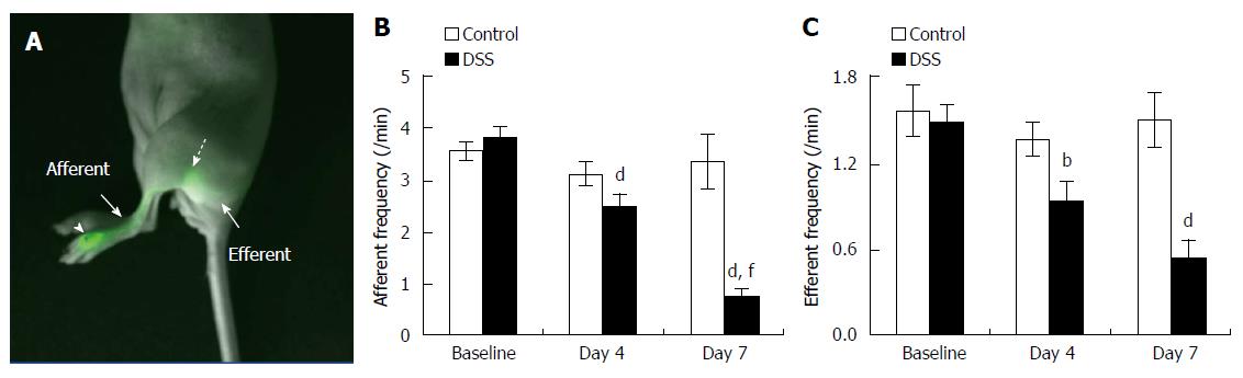
Figure 5 Lymphatic contractile function in the skin of mice with dextran sulfate sodium-induced acute colitis.
Overlay of fluorescent and white light images showing lymphatic drainage of indocyanine green (ICG) from the foot, where ICG was injected (arrowhead), to the popliteal LN (broken arrow) via the popliteal afferent lymphatic vessel (A). The quantification of lymphatic contractility in the popliteal afferent (B) and efferent (C) lymphatic vessels in the foot of mice prior to 4, and 7 d after DSS alone (n = 21). Data represent mean ± SE. bP < 0.01, dP < 0.001 vs baseline; fP < 0.001 vs day 4.
- Citation: Agollah GD, Wu G, Peng HL, Kwon S. Dextran sulfate sodium-induced acute colitis impairs dermal lymphatic function in mice. World J Gastroenterol 2015; 21(45): 12767-12777
- URL: https://www.wjgnet.com/1007-9327/full/v21/i45/12767.htm
- DOI: https://dx.doi.org/10.3748/wjg.v21.i45.12767