
















©The Author(s) 2015.
World J Gastroenterol. Dec 7, 2015; 21(45): 12767-12777
Published online Dec 7, 2015. doi: 10.3748/wjg.v21.i45.12767
Published online Dec 7, 2015. doi: 10.3748/wjg.v21.i45.12767
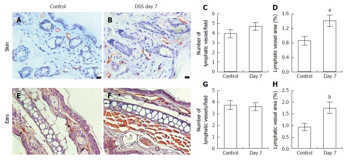
Figure 4 Immunohistochemical assessment of lymphatic vessels using antibodies to LYVE-1.
In the skin (A) and ears (E, arrows) of control (n = 5 skin; n = 7 ear) and mice with dextran sulfate sodium (DSS)-induced acute colitis (B, F; n = 6 skin; n = 9 ears). Computer-assisted image analysis showed no difference in the number of lymphatic vessels per field (C, G) but increased lymphatic vessel area in both skin and ears (D, H) compared to control mice. Data presented as mean ± SE. aP < 0.05, bP < 0.01 vs control. Scale bar = 100 μm (C, D).
- Citation: Agollah GD, Wu G, Peng HL, Kwon S. Dextran sulfate sodium-induced acute colitis impairs dermal lymphatic function in mice. World J Gastroenterol 2015; 21(45): 12767-12777
- URL: https://www.wjgnet.com/1007-9327/full/v21/i45/12767.htm
- DOI: https://dx.doi.org/10.3748/wjg.v21.i45.12767