
















©The Author(s) 2015.
World J Gastroenterol. Aug 28, 2015; 21(32): 9544-9553
Published online Aug 28, 2015. doi: 10.3748/wjg.v21.i32.9544
Published online Aug 28, 2015. doi: 10.3748/wjg.v21.i32.9544
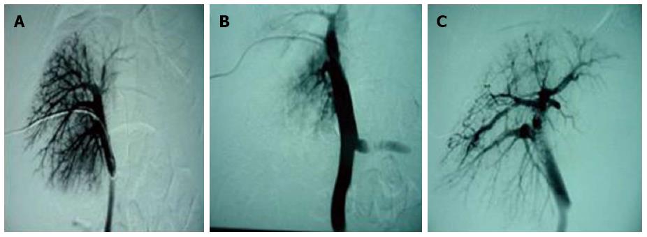
Figure 2 Portal vein variation.
A: Direct portal phlebography of experimental group 2 before perfusion showed the portal venous branches clearly; B: Portal phlebography immediately after perfusion showed a significant reduction in small portal venous branches and a widened main portal vein; C: Portal phlebography at 6 wk after perfusion showed that a large number of irregular bypass branches of intrahepatic portal veins were formed, which communicated directly with hepatic veins, and the main portal vein was still wider than that before perfusion.
- Citation: Wang L, He FL, Liu FQ, Yue ZD, Zhao HW. Establishment of a hepatic cirrhosis and portal hypertension model by hepatic arterial perfusion with 80% alcohol. World J Gastroenterol 2015; 21(32): 9544-9553
- URL: https://www.wjgnet.com/1007-9327/full/v21/i32/9544.htm
- DOI: https://dx.doi.org/10.3748/wjg.v21.i32.9544