
















©The Author(s) 2015.
World J Gastroenterol. Jan 21, 2015; 21(3): 1024-1027
Published online Jan 21, 2015. doi: 10.3748/wjg.v21.i3.1024
Published online Jan 21, 2015. doi: 10.3748/wjg.v21.i3.1024
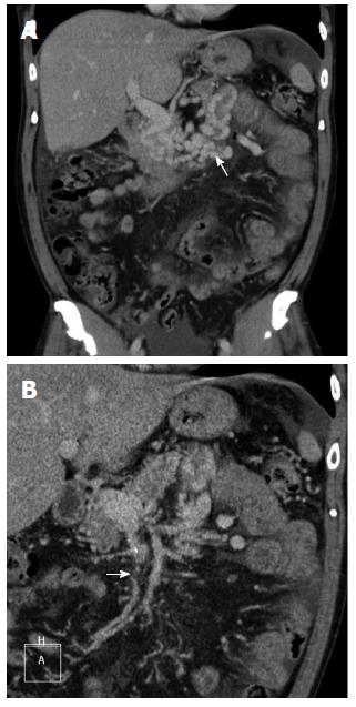
Figure 1 Computed tomographic scan reveals vascular tufts around the proximal jejunum (A, arrow), and thrombi are visible as hypodense lesions in the contrasted superior mesenteric vein (B, arrow).
- Citation: Hsu WF, Tsang YM, Teng CJ, Chung CS. Protein C deficiency related obscure gastrointestinal bleeding treated by enteroscopy and anticoagulant therapy. World J Gastroenterol 2015; 21(3): 1024-1027
- URL: https://www.wjgnet.com/1007-9327/full/v21/i3/1024.htm
- DOI: https://dx.doi.org/10.3748/wjg.v21.i3.1024