
















©The Author(s) 2015.
World J Gastroenterol. Jul 21, 2015; 21(27): 8452-8457
Published online Jul 21, 2015. doi: 10.3748/wjg.v21.i27.8452
Published online Jul 21, 2015. doi: 10.3748/wjg.v21.i27.8452
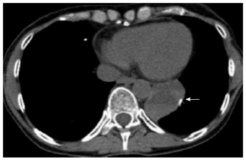
Figure 1 Non-contrast chest computed tomography demonstrates a round mass in the left inferior paramediastinal-pleural region with heterogeneous density (arrow).
There is surgical hyperdense material seen at the margin of the round mass and right anterior lung region, which was from prior metastases resection.
- Citation: Akpinar B, Obuch J, Fukami N, Pokharel SS. Unusual presentation of a pancreatic cyst resulting from osteosarcoma metastasis. World J Gastroenterol 2015; 21(27): 8452-8457
- URL: https://www.wjgnet.com/1007-9327/full/v21/i27/8452.htm
- DOI: https://dx.doi.org/10.3748/wjg.v21.i27.8452