
















©The Author(s) 2015.
World J Gastroenterol. Jul 14, 2015; 21(26): 8215-8220
Published online Jul 14, 2015. doi: 10.3748/wjg.v21.i26.8215
Published online Jul 14, 2015. doi: 10.3748/wjg.v21.i26.8215
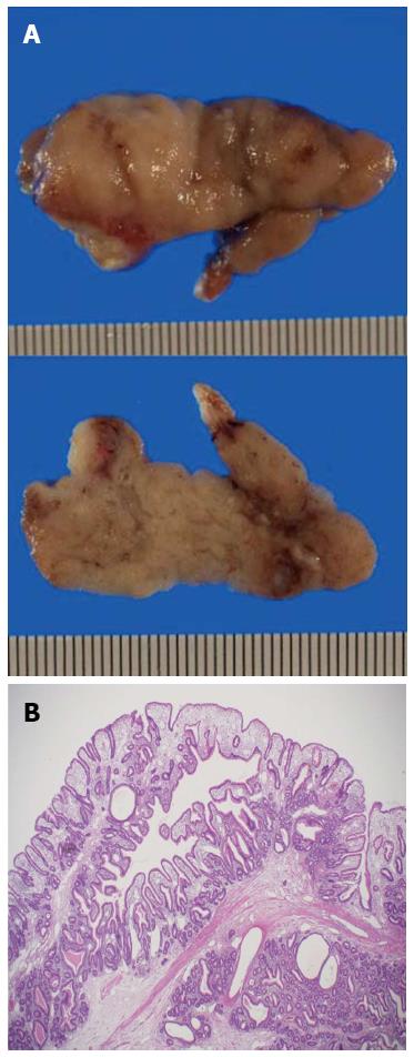
Figure 4 Histopathologic findings.
A: View of the surgical specimen, showing that it was 4.8 cm × 1.9 cm × 1.8 cm in diameter with lobular surfaces, which were epithelialized with intestinal type mucosa; B: Histopathologic examination showed features suggesting a hamartoma. For example, the polyp contained irregular hyperplastic crypts with focal cystic dilatation and branching bundles of smooth muscle extending from the muscularis mucosae. The epithelium was hyperplastic.
- Citation: Suzuki K, Higuchi H, Shimizu S, Nakano M, Serizawa H, Morinaga S. Endoscopic snare papillectomy for a solitary Peutz-Jeghers-type polyp in the duodenum with ingrowth into the common bile duct: Case report. World J Gastroenterol 2015; 21(26): 8215-8220
- URL: https://www.wjgnet.com/1007-9327/full/v21/i26/8215.htm
- DOI: https://dx.doi.org/10.3748/wjg.v21.i26.8215