
















©The Author(s) 2015.
World J Gastroenterol. Jul 14, 2015; 21(26): 8163-8169
Published online Jul 14, 2015. doi: 10.3748/wjg.v21.i26.8163
Published online Jul 14, 2015. doi: 10.3748/wjg.v21.i26.8163
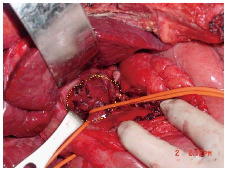
Figure 3 Anatomy of caudate lobe fossa and inferior vena cava.
Caudate lobe fossa (circumscribed by yellow dotted zone) and inferior vena cava (arrow) following removal of caudate lobe.
- Citation: Hong DF, Liu YB, Peng SY, Pang JZ, Wang ZF, Cheng J, Shen GL, Zhang YB. Management of hepatocellular carcinoma rupture in the caudate lobe. World J Gastroenterol 2015; 21(26): 8163-8169
- URL: https://www.wjgnet.com/1007-9327/full/v21/i26/8163.htm
- DOI: https://dx.doi.org/10.3748/wjg.v21.i26.8163