
















©The Author(s) 2015.
World J Gastroenterol. Jun 28, 2015; 21(24): 7589-7593
Published online Jun 28, 2015. doi: 10.3748/wjg.v21.i24.7589
Published online Jun 28, 2015. doi: 10.3748/wjg.v21.i24.7589
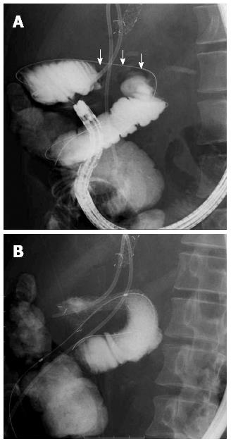
Figure 1 A contrast medium was injected and the stricture of the afferent loop was observed (arrow) (A), a guidewire was passed through the stenosis and placed under the single balloon enteroscope, placement of the enteral metallic stent along the guidewire in the overtube (B).
- Citation: Nakahara K, Okuse C, Matsumoto N, Suetani K, Morita R, Michikawa Y, Ozawa SI, Hosoya K, Kobayashi S, Otsubo T, Itoh F. Enteral metallic stenting by balloon enteroscopy for obstruction of surgically reconstructed intestine. World J Gastroenterol 2015; 21(24): 7589-7593
- URL: https://www.wjgnet.com/1007-9327/full/v21/i24/7589.htm
- DOI: https://dx.doi.org/10.3748/wjg.v21.i24.7589