
















©The Author(s) 2015.
World J Gastroenterol. Jun 7, 2015; 21(21): 6754-6758
Published online Jun 7, 2015. doi: 10.3748/wjg.v21.i21.6754
Published online Jun 7, 2015. doi: 10.3748/wjg.v21.i21.6754
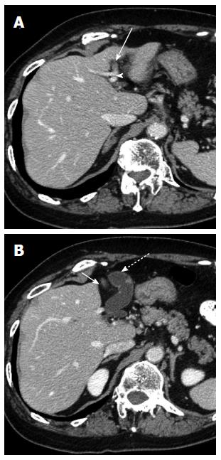
Figure 1 Computed tomography examination reveals a left-sided gallbladder.
A: The round ligament (arrow) is connected to the left portal umbilical portion (arrow head); B: The gallbladder (dotted arrow) is located to the left side of the round ligament (arrow) and the middle hepatic vein, and attached to the left lateral section of the liver.
- Citation: Ishii H, Noguchi A, Onishi M, Takao K, Maruyama T, Taiyoh H, Araki Y, Shimizu T, Izumi H, Tani N, Yamaguchi M, Yamane T. True left-sided gallbladder with variations of bile duct and cholecystic vein. World J Gastroenterol 2015; 21(21): 6754-6758
- URL: https://www.wjgnet.com/1007-9327/full/v21/i21/6754.htm
- DOI: https://dx.doi.org/10.3748/wjg.v21.i21.6754