
















©The Author(s) 2015.
World J Gastroenterol. Apr 21, 2015; 21(15): 4509-4516
Published online Apr 21, 2015. doi: 10.3748/wjg.v21.i15.4509
Published online Apr 21, 2015. doi: 10.3748/wjg.v21.i15.4509
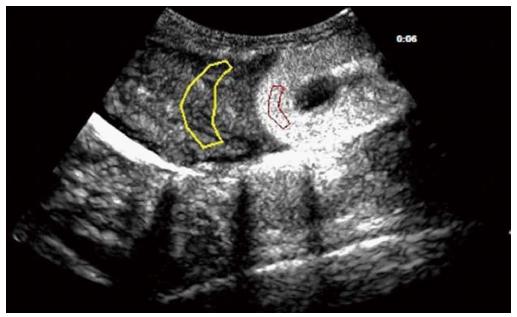
Figure 1 Contrast-enhanced ultrasonography of the right lobe of the liver and the upper pole of the kidney in a sagittal view.
The region defined in yellow is the region of interest (ROI) of the hepatic parenchyma, with the region in red being the ROI of the renal cortex.
- Citation: Zhai L, Qiu LY, Zu Y, Yan Y, Ren XZ, Zhao JF, Liu YJ, Liu JB, Qian LX. Contrast-enhanced ultrasound for quantitative assessment of portal pressure in canine liver fibrosis. World J Gastroenterol 2015; 21(15): 4509-4516
- URL: https://www.wjgnet.com/1007-9327/full/v21/i15/4509.htm
- DOI: https://dx.doi.org/10.3748/wjg.v21.i15.4509