
















©The Author(s) 2015.
World J Gastroenterol. Mar 28, 2015; 21(12): 3579-3586
Published online Mar 28, 2015. doi: 10.3748/wjg.v21.i12.3579
Published online Mar 28, 2015. doi: 10.3748/wjg.v21.i12.3579
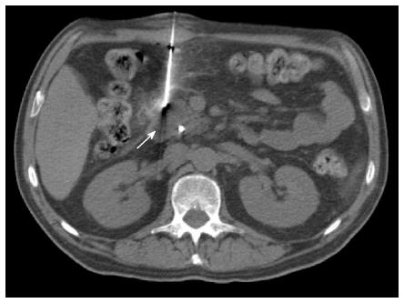
Figure 1 Percutaneous computed tomography-guided core needle biopsy of a pancreatic lesion using direct access.
Non-enhanced axial computed tomography of the upper abdomen showing an anteriorly inserted coaxial needle (17 G) that is placed directly over the lesion located at the head of the pancreas (arrow).
- Citation: Tyng CJ, Almeida MFA, Barbosa PN, Bitencourt AG, Berg JAA, Maciel MS, Coimbra FJ, Schiavon LHO, Begnami MD, Guimarães MD, Zurstrassen CE, Chojniak R. Computed tomography-guided percutaneous core needle biopsy in pancreatic tumor diagnosis. World J Gastroenterol 2015; 21(12): 3579-3586
- URL: https://www.wjgnet.com/1007-9327/full/v21/i12/3579.htm
- DOI: https://dx.doi.org/10.3748/wjg.v21.i12.3579