
















©The Author(s) 2015.
World J Gastroenterol. Mar 21, 2015; 21(11): 3429-3434
Published online Mar 21, 2015. doi: 10.3748/wjg.v21.i11.3429
Published online Mar 21, 2015. doi: 10.3748/wjg.v21.i11.3429
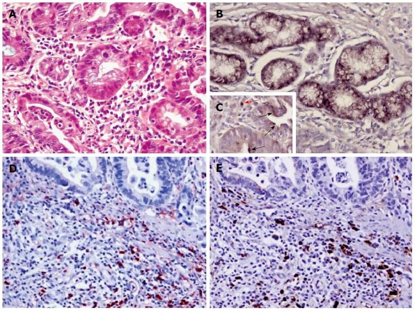
Figure 3 Histological findings of the endoscopic biopsy specimen from the pylorus.
HE staining shows a moderately differentiated gastric adenocarcinoma with abundant infiltration of lymphoplasmacytes and eosinophils in stroma (A). Immunostaining reveals Helicobacter pylori in gastric epithelial cells (B) or cancer cells (black arrow, C) or mesenchymal cells (red arrow, C), IgG4-positive (D) or IgG-positive plasma cells (E) in the cancer stroma. Original magnification × 400 (A, B and C), × 200 (D and E).
-
Citation: Li M, Zhou Q, Yang K, Brigstock DR, Zhang L, Xiu M, Sun L, Gao RP. Rare case of
Helicobacter pylori -positive multiorgan IgG4-related disease and gastric cancer. World J Gastroenterol 2015; 21(11): 3429-3434 - URL: https://www.wjgnet.com/1007-9327/full/v21/i11/3429.htm
- DOI: https://dx.doi.org/10.3748/wjg.v21.i11.3429