
















©2014 Baishideng Publishing Group Inc.
World J Gastroenterol. Oct 21, 2014; 20(39): 14495-14499
Published online Oct 21, 2014. doi: 10.3748/wjg.v20.i39.14495
Published online Oct 21, 2014. doi: 10.3748/wjg.v20.i39.14495
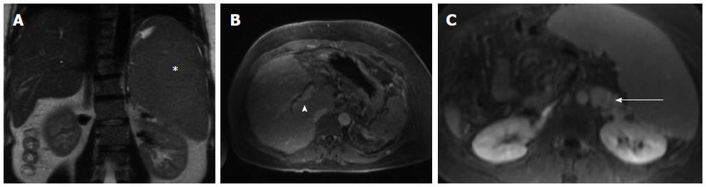
Figure 1 Magnetic resonance imaging - abdomen from two months prior to embolization.
Massive splenomegaly is shown (A, asterisk) with associated cavernous transformation of the portal vein (B, arrow head) and large splenorenal varices (C, arrow).
- Citation: Gianotti R, Charles H, Hymes K, Chandarana H, Sigal S. Treatment of gastric varices with partial splenic embolization in a patient with portal vein thrombosis and a myeloproliferative disorder. World J Gastroenterol 2014; 20(39): 14495-14499
- URL: https://www.wjgnet.com/1007-9327/full/v20/i39/14495.htm
- DOI: https://dx.doi.org/10.3748/wjg.v20.i39.14495