
















©2014 Baishideng Publishing Group Inc.
World J Gastroenterol. Aug 28, 2014; 20(32): 11287-11296
Published online Aug 28, 2014. doi: 10.3748/wjg.v20.i32.11287
Published online Aug 28, 2014. doi: 10.3748/wjg.v20.i32.11287
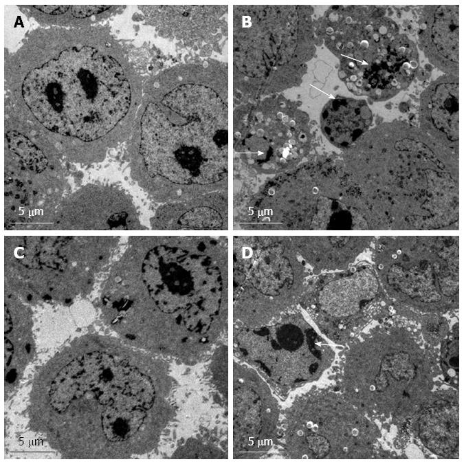
Figure 5 Transmission electron microscopy.
The ultrastructural morphology of control (A) and RNAi (B) of Hep G2 cells. The ultrastructural morphology of control (C) and RNAi (D) Bel7402 cells. Nuclear fragmentation and apoptotic bodies are indicated by arrows.
-
Citation: Zhang YL, Zhang YC, Han W, Li YM, Wang GN, Yuan S, Wei FX, Wang JF, Jiang JJ, Zhang YW. Effect of
GP73 silencing on proliferation and apoptosis in hepatocellular cancer. World J Gastroenterol 2014; 20(32): 11287-11296 - URL: https://www.wjgnet.com/1007-9327/full/v20/i32/11287.htm
- DOI: https://dx.doi.org/10.3748/wjg.v20.i32.11287