
















©2014 Baishideng Publishing Group Inc.
World J Gastroenterol. Aug 7, 2014; 20(29): 9699-9715
Published online Aug 7, 2014. doi: 10.3748/wjg.v20.i29.9699
Published online Aug 7, 2014. doi: 10.3748/wjg.v20.i29.9699
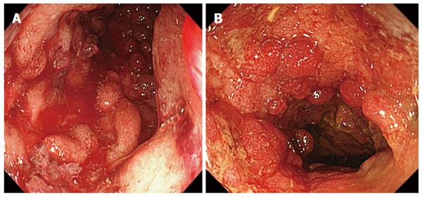
Figure 9 Colonoscopy images showing deep and extensive colonic lesions together with inflammatory polyps and contact bleeding.
Typical colonoscopic images from patients with severely damaged mucosal tissue (A), granulocyte/monocyte apheresis non-responders (B). However, most patients with the entry mucosal damage seen in this figure are medication refractory and unlikely to respond to granulocyte and monocyte apheresis, some opt for colectomy.
- Citation: Saniabadi AR, Tanaka T, Ohmori T, Sawada K, Yamamoto T, Hanai H. Treating inflammatory bowel disease by adsorptive leucocytapheresis: A desire to treat without drugs. World J Gastroenterol 2014; 20(29): 9699-9715
- URL: https://www.wjgnet.com/1007-9327/full/v20/i29/9699.htm
- DOI: https://dx.doi.org/10.3748/wjg.v20.i29.9699