
















©2014 Baishideng Publishing Group Inc.
World J Gastroenterol. Jun 7, 2014; 20(21): 6675-6679
Published online Jun 7, 2014. doi: 10.3748/wjg.v20.i21.6675
Published online Jun 7, 2014. doi: 10.3748/wjg.v20.i21.6675
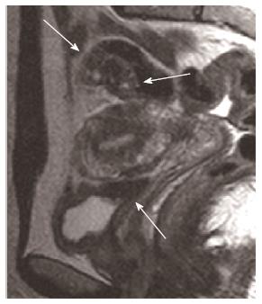
Figure 2 Magnetic resonance imaging shows two hypointense nodular implants of endometriosis infiltrating the proximal sigmoid wall and the utero-vesical fold, with the latter retrospectively recognized after the surgery (arrows).
The presence of high signal intensity foci within the nodules is due to hemorrhagic foci.
- Citation: Cacciato Insilla A, Granai M, Gallippi G, Giusti P, Giusti S, Guadagni S, Morelli L, Campani D. Deep endometriosis with pericolic lymph node involvement: A case report and literature review. World J Gastroenterol 2014; 20(21): 6675-6679
- URL: https://www.wjgnet.com/1007-9327/full/v20/i21/6675.htm
- DOI: https://dx.doi.org/10.3748/wjg.v20.i21.6675