
















©2014 Baishideng Publishing Group Inc.
World J Gastroenterol. Jun 7, 2014; 20(21): 6615-6619
Published online Jun 7, 2014. doi: 10.3748/wjg.v20.i21.6615
Published online Jun 7, 2014. doi: 10.3748/wjg.v20.i21.6615
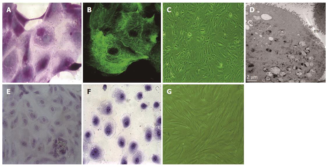
Figure 1 Periodic acid-Schiff, cytokeratin-18 and toluidine blue staining and cell morphology.
A: Cytoplasm of normal human gastric mucosal epithelial cells (nhGMECs) was stained purple with periodic acid-Schiff (PAS), and contained neutral mucin granules, magnification × 1000; B: nhGMEC network-structure staining with an antibody against cytokeratin (CK)-18 demonstrates the presence of CK-18; C: Phase-contrast micrograph of nhGMECs after 4 d of culture, magnification × 100; D: Transmission electron microscopy revealed the presence of microvilli (mv) and secretary granules (sg) in nhGMECs, magnification × 1000; E: nhGMECs were detected by toluidine blue staining, and nuclei (light color) were observed, magnification × 400; F: SGC-7901 cells were detected by toluidine blue staining, and nuclei with multiple nucleoli (deep color) were observed, magnification × 400; G: Phase-contrast micrograph of nhGMFs after 13 d of culture, magnification × 100.
- Citation: Cheng YB, Guo LP, Yao P, Ning XY, Aerken G, Fang DC. Telomerase and hTERT: Can they serve as markers for gastric cancer diagnosis? World J Gastroenterol 2014; 20(21): 6615-6619
- URL: https://www.wjgnet.com/1007-9327/full/v20/i21/6615.htm
- DOI: https://dx.doi.org/10.3748/wjg.v20.i21.6615