
















©2014 Baishideng Publishing Group Co.
World J Gastroenterol. May 7, 2014; 20(17): 5157-5164
Published online May 7, 2014. doi: 10.3748/wjg.v20.i17.5157
Published online May 7, 2014. doi: 10.3748/wjg.v20.i17.5157
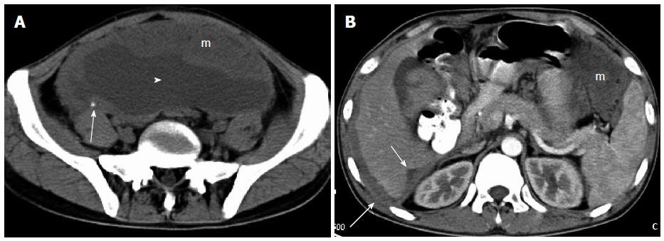
Figure 4 A 24-year-old man with faint abdominal pain.
A: Plain computed tomography (CT) demonstrated a large, wavy, omental soft-tissue mass (m) with foci of calcification (arrow), and massive ascites (arrowhead); B: Enhanced CT showed diffuse thickening of the perihepatic parietal peritoneum with liver infiltration (arrows), and omental mass (m). All of the tumor tissues presented with slight uniform enhancement.
- Citation: Shen XZ, Zhao JG, Wu JJ, Liu F. Clinical and computed tomography features of adult abdominopelvic desmoplastic small round cell tumor. World J Gastroenterol 2014; 20(17): 5157-5164
- URL: https://www.wjgnet.com/1007-9327/full/v20/i17/5157.htm
- DOI: https://dx.doi.org/10.3748/wjg.v20.i17.5157