
















©2014 Baishideng Publishing Group Co.
World J Gastroenterol. Apr 28, 2014; 20(16): 4712-4717
Published online Apr 28, 2014. doi: 10.3748/wjg.v20.i16.4712
Published online Apr 28, 2014. doi: 10.3748/wjg.v20.i16.4712
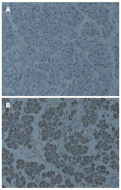
Figure 3 Immunohistochemical staining of Ptch1 in pancreas (× 200).
A: Normal rats; B: Chronic pancreatitis rats. Significantly higher expression of Ptch1 is seen in Figure B, and Ptch1 expression was mainly restricted to ductal, acinar, or islet compartments of the pancreas.
- Citation: Wang LW, Lin H, Lu Y, Xia W, Gao J, Li ZS. Sonic hedgehog expression in a rat model of chronic pancreatitis. World J Gastroenterol 2014; 20(16): 4712-4717
- URL: https://www.wjgnet.com/1007-9327/full/v20/i16/4712.htm
- DOI: https://dx.doi.org/10.3748/wjg.v20.i16.4712