
















©2014 Baishideng Publishing Group Co.
World J Gastroenterol. Apr 14, 2014; 20(14): 4110-4114
Published online Apr 14, 2014. doi: 10.3748/wjg.v20.i14.4110
Published online Apr 14, 2014. doi: 10.3748/wjg.v20.i14.4110
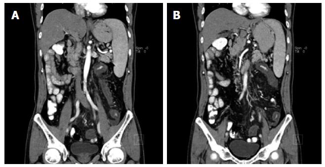
Figure 1 Magnetic resonance imaging of the abdomen and pelvic showed a stenosis of the mesenteric inferior artery branch (A) and thickening of the mesenteric inferior artery branch walls (B).
The image shows a diffuse lesion of the left colon suggesting ischaemic injury.
- Citation: Yang XN, Ye ZS, Fan YY, Hu YQ. Prolonged small vessel vasculitis with colon mucosal inflammation as first manifestations of Behçet’s disease. World J Gastroenterol 2014; 20(14): 4110-4114
- URL: https://www.wjgnet.com/1007-9327/full/v20/i14/4110.htm
- DOI: https://dx.doi.org/10.3748/wjg.v20.i14.4110