
















©2013 Baishideng Publishing Group Co.
World J Gastroenterol. Feb 28, 2013; 19(8): 1230-1238
Published online Feb 28, 2013. doi: 10.3748/wjg.v19.i8.1230
Published online Feb 28, 2013. doi: 10.3748/wjg.v19.i8.1230
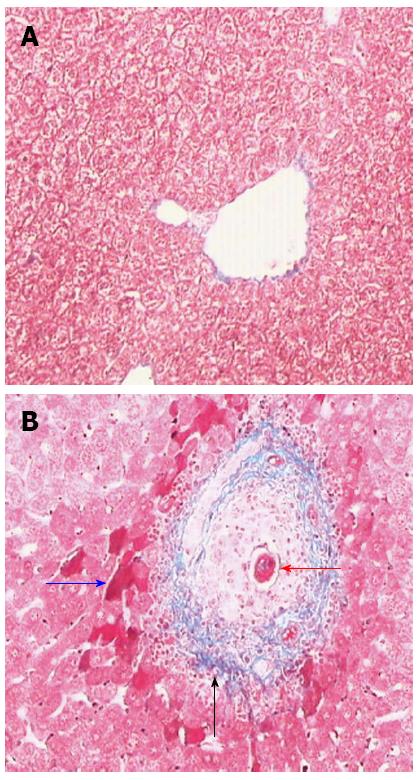
Figure 1 Masson staining of 6 wk post-infect mice livers (× 200).
A: Normal mice; B: Infected mice, the red arrow indicated the eggs deposited in the vein, the blue represented acidophilic necrosis, and the black was the collagen fiber deposited around the vein.
- Citation: Liu P, Wang M, Lu XD, Zhang SJ, Tang WX. Schistosoma japonicum egg antigen up-regulates fibrogenesis and inhibits proliferation in primary hepatic stellate cells in a concentration-dependent manner. World J Gastroenterol 2013; 19(8): 1230-1238
- URL: https://www.wjgnet.com/1007-9327/full/v19/i8/1230.htm
- DOI: https://dx.doi.org/10.3748/wjg.v19.i8.1230