
















©2013 Baishideng Publishing Group Co.
World J Gastroenterol. Nov 28, 2013; 19(44): 8156-8159
Published online Nov 28, 2013. doi: 10.3748/wjg.v19.i44.8156
Published online Nov 28, 2013. doi: 10.3748/wjg.v19.i44.8156
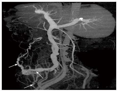
Figure 2 Three-dimensional reconstruction showed the ectopic varices (black arrow) at the ileal conduit stoma fed by the superior mesenteric vein (white arrow) and communicated to the paraumbilical vein (white arrow) and femoral vein (white arrow).
- Citation: Yao DH, Luo XF, Zhou B, Li X. Ileal conduit stomal variceal bleeding managed by endovascular embolization. World J Gastroenterol 2013; 19(44): 8156-8159
- URL: https://www.wjgnet.com/1007-9327/full/v19/i44/8156.htm
- DOI: https://dx.doi.org/10.3748/wjg.v19.i44.8156