
















©2013 Baishideng Publishing Group Co.
World J Gastroenterol. Jul 28, 2013; 19(28): 4616-4623
Published online Jul 28, 2013. doi: 10.3748/wjg.v19.i28.4616
Published online Jul 28, 2013. doi: 10.3748/wjg.v19.i28.4616
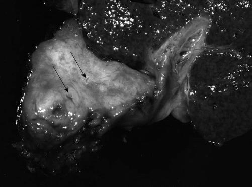
Figure 1 Gross appearance of the hilar neoplasm.
The resected specimen showed a firm, tan-grey tumor measuring 5.0-cm in greatest dimension with extensive involvement of the perihilar bile ducts and surrounding soft tissue. Some of the small ducts and vessels within the lesion showed severe luminal narrowing (arrows).
- Citation: Sasatomi E, Nalesnik MA, Marsh JW. Neuroendocrine carcinoma of the extrahepatic bile duct: Case report and literature review. World J Gastroenterol 2013; 19(28): 4616-4623
- URL: https://www.wjgnet.com/1007-9327/full/v19/i28/4616.htm
- DOI: https://dx.doi.org/10.3748/wjg.v19.i28.4616