
















©2013 Baishideng Publishing Group Co.
World J Gastroenterol. May 28, 2013; 19(20): 3161-3164
Published online May 28, 2013. doi: 10.3748/wjg.v19.i20.3161
Published online May 28, 2013. doi: 10.3748/wjg.v19.i20.3161
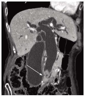
Figure 1 Computed tomography of the bile duct.
Computed tomography revealed dilatation of the bile duct and an elevated lesion (arrow) at the bottom of the lower bile duct.
- Citation: Onishi I, Kitagawa H, Harada K, Maruzen S, Sakai S, Makino I, Hayashi H, Nakagawara H, Tajima H, Takamura H, Tani T, Kayahara M, Ikeda H, Ohta T, Nakanuma Y. Intraductal papillary neoplasm of the bile duct accompanying biliary mixed adenoneuroendocrine carcinoma. World J Gastroenterol 2013; 19(20): 3161-3164
- URL: https://www.wjgnet.com/1007-9327/full/v19/i20/3161.htm
- DOI: https://dx.doi.org/10.3748/wjg.v19.i20.3161