
















©2013 Baishideng Publishing Group Co.
World J Gastroenterol. Mar 28, 2013; 19(12): 1919-1929
Published online Mar 28, 2013. doi: 10.3748/wjg.v19.i12.1919
Published online Mar 28, 2013. doi: 10.3748/wjg.v19.i12.1919
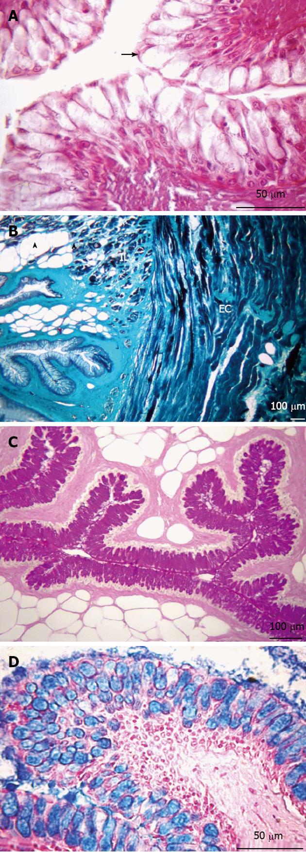
Figure 2 Transversal sections of the esophagus.
A: Non-keratinized stratified squamous epithelium (arrow). Hematoxylin and eosin stain; B: Presence of adipose tissue (arrowheads) in the submucosa layer. Muscular layer formed by two sub-layers, internal longitudinal (IL) and external circular (EC). Gomori’s trichrome stain; C: Presence of neutral glycoconjugates (GCs). Periodic acid-Schiff stain; D: Acid GCs. Alcian blue stain.
-
Citation: Vieira-Lopes DA, Pinheiro NL, Sales A, Ventura A, Araújo FG, Gomes ID, Nascimento AA. Immunohistochemical study of the digestive tract of
Oligosarcus hepsetus . World J Gastroenterol 2013; 19(12): 1919-1929 - URL: https://www.wjgnet.com/1007-9327/full/v19/i12/1919.htm
- DOI: https://dx.doi.org/10.3748/wjg.v19.i12.1919