
















©2013 Baishideng Publishing Group Co.
World J Gastroenterol. Mar 21, 2013; 19(11): 1841-1844
Published online Mar 21, 2013. doi: 10.3748/wjg.v19.i11.1841
Published online Mar 21, 2013. doi: 10.3748/wjg.v19.i11.1841
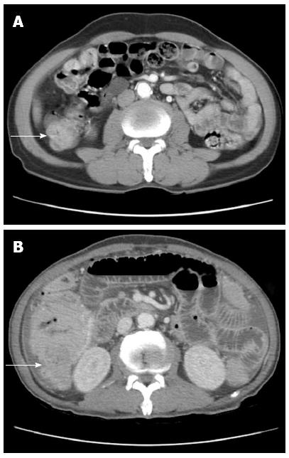
Figure 1 An abdominopelvic computed tomography shows segmental wall thickening of distal ascending with nonspecific multiple small lymphnodes along ileocolic vessels.
Large irregular mass (A) in right ascending colon along hepatic flexure (B).
- Citation: Kim JB, Kim SH, Cho YK, Ahn SB, Jo YJ, Park YS, Lee JH, Kim DH, Lee H, Jung YY. A case of colon perforation due to enteropathy-associated T-cell lymphoma. World J Gastroenterol 2013; 19(11): 1841-1844
- URL: https://www.wjgnet.com/1007-9327/full/v19/i11/1841.htm
- DOI: https://dx.doi.org/10.3748/wjg.v19.i11.1841