
















©2012 Baishideng Publishing Group Co.
World J Gastroenterol. Jul 14, 2012; 18(26): 3379-3388
Published online Jul 14, 2012. doi: 10.3748/wjg.v18.i26.3379
Published online Jul 14, 2012. doi: 10.3748/wjg.v18.i26.3379
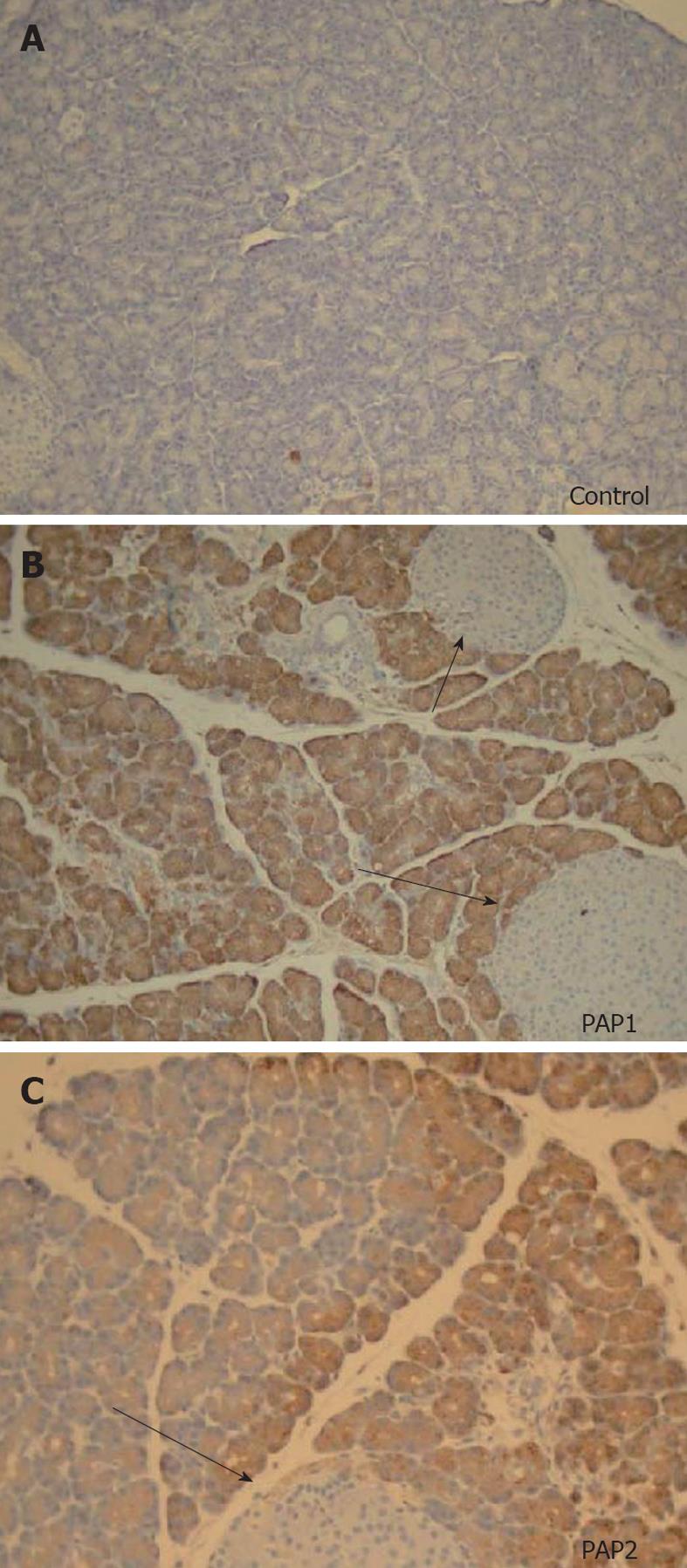
Figure 6 Immunohistochemistry for pancreatitis-associated proteins (40×) in a young control animal without pancreatitis (A) compared to pancreatitis-associated protein 1 (B) and pancreatitis-associated protein 2 (C) in young animals 24 h after sodium taurocholate pancreatitis.
No staining is seen in controls, uniform brown colored staining is seen for pancreatitis-associated protein (PAP)1 (B). Preparation of the slides was done side by side. A different, heterogenous pattern of staining is seen for PAP2 (C). Note that islets of Langerhans (arrows) do not stain at all for PAPs in normal animals or animals with acute pancreatitis.
- Citation: Fu S, Stanek A, Mueller CM, Brown NA, Huan C, Bluth MH, Zenilman ME. Acute pancreatitis in aging animals: Loss of pancreatitis-associated protein protection? World J Gastroenterol 2012; 18(26): 3379-3388
- URL: https://www.wjgnet.com/1007-9327/full/v18/i26/3379.htm
- DOI: https://dx.doi.org/10.3748/wjg.v18.i26.3379