
















©2012 Baishideng Publishing Group Co.
World J Gastroenterol. Mar 28, 2012; 18(12): 1328-1338
Published online Mar 28, 2012. doi: 10.3748/wjg.v18.i12.1328
Published online Mar 28, 2012. doi: 10.3748/wjg.v18.i12.1328
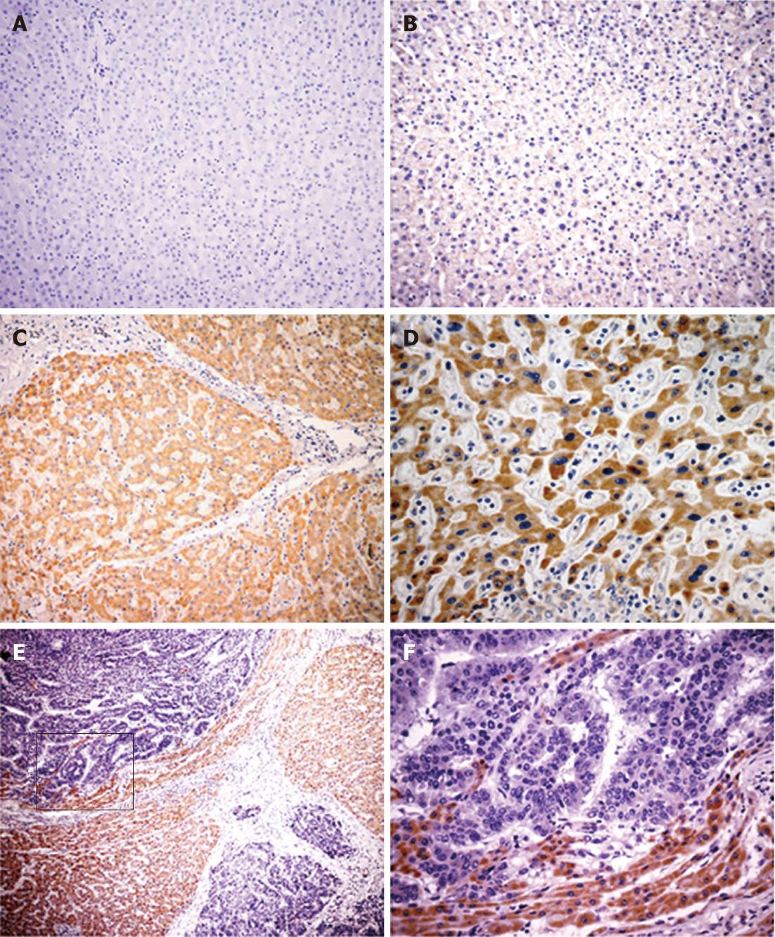
Figure 2 Immunohistochemical staining for Ror2 in hepatocellular carcinoma.
Patient-matched normal liver cells showed negative (A) or weak expression (B) of receptor tyrosine kinase-like orphan receptor 2 (Ror2). Liver cirrhosis cells (C) and dysplastic liver cells (D) exhibited strong positive immunostaining for Ror2. Tumor cells (E, F) showing negative Ror2 staining in hepatocytes, while strong cytoplasmic staining is seen in adjacent nontumorous cells. Original magnification, 200 × in A; 400 × in B and D; 100 × in C.
- Citation: Geng M, Cao YC, Chen YJ, Jiang H, Bi LQ, Liu XH. Loss of Wnt5a and Ror2 protein in hepatocellular carcinoma associated with poor prognosis. World J Gastroenterol 2012; 18(12): 1328-1338
- URL: https://www.wjgnet.com/1007-9327/full/v18/i12/1328.htm
- DOI: https://dx.doi.org/10.3748/wjg.v18.i12.1328