
















©2011 Baishideng Publishing Group Co.
World J Gastroenterol. Nov 14, 2011; 17(42): 4734-4738
Published online Nov 14, 2011. doi: 10.3748/wjg.v17.i42.4734
Published online Nov 14, 2011. doi: 10.3748/wjg.v17.i42.4734
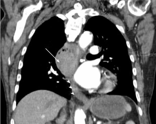
Figure 1 Coronal reformat of a contrast-enhanced chest computed tomography demonstrates a solid enhancing mass with soft tissue attenuation in the middle third of the esophagus (arrow) causing obstruction and proximal dilatation.
The distal third of the esophagus (arrowheads) is unremarkable.
- Citation: Machado J, Ministro P, Araújo R, Cancela E, Castanheira A, Silva A. Primary malignant melanoma of the esophagus: A case report. World J Gastroenterol 2011; 17(42): 4734-4738
- URL: https://www.wjgnet.com/1007-9327/full/v17/i42/4734.htm
- DOI: https://dx.doi.org/10.3748/wjg.v17.i42.4734