
















©2011 Baishideng Publishing Group Co.
World J Gastroenterol. Jan 28, 2011; 17(4): 433-443
Published online Jan 28, 2011. doi: 10.3748/wjg.v17.i4.433
Published online Jan 28, 2011. doi: 10.3748/wjg.v17.i4.433
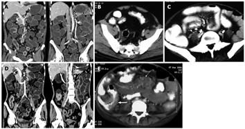
Figure 3 Findings on computed tomography.
A: Computed tomography (CT) enteroclysis with negative oral contrast showing mural thickening of ileum with skip areas and sparing of cecum in a patient with Crohn's disease (CD); B: Contrast-enhanced CT scan (CECT) showing asymmetrical mural thickening in ileal loop with deep ulcerations. Note fibro-fatty proliferation of mesentery; C: CECT in another patient with CD showing mesenteric vascular engorgement (Comb sign) with fibro-fatty proliferation of the mesentery; D: CT enteroclysis with negative oral contrast showing contiguous mural thickening of the terminal ileum and cecum in a patient with tuberculosis (TB); E: CECT in a patient with TB showing mural thickening of the terminal ileum and cecum (thick arrow) with multiple enlarged mesenteric lymph nodes showing central hypoattenuating and peripherally enhancing rims (thin arrow).
- Citation: Pulimood AB, Amarapurkar DN, Ghoshal U, Phillip M, Pai CG, Reddy DN, Nagi B, Ramakrishna BS. Differentiation of Crohn’s disease from intestinal tuberculosis in India in 2010. World J Gastroenterol 2011; 17(4): 433-443
- URL: https://www.wjgnet.com/1007-9327/full/v17/i4/433.htm
- DOI: https://dx.doi.org/10.3748/wjg.v17.i4.433