
















©2011 Baishideng Publishing Group Co.
World J Gastroenterol. Sep 21, 2011; 17(35): 4038-4043
Published online Sep 21, 2011. doi: 10.3748/wjg.v17.i35.4038
Published online Sep 21, 2011. doi: 10.3748/wjg.v17.i35.4038
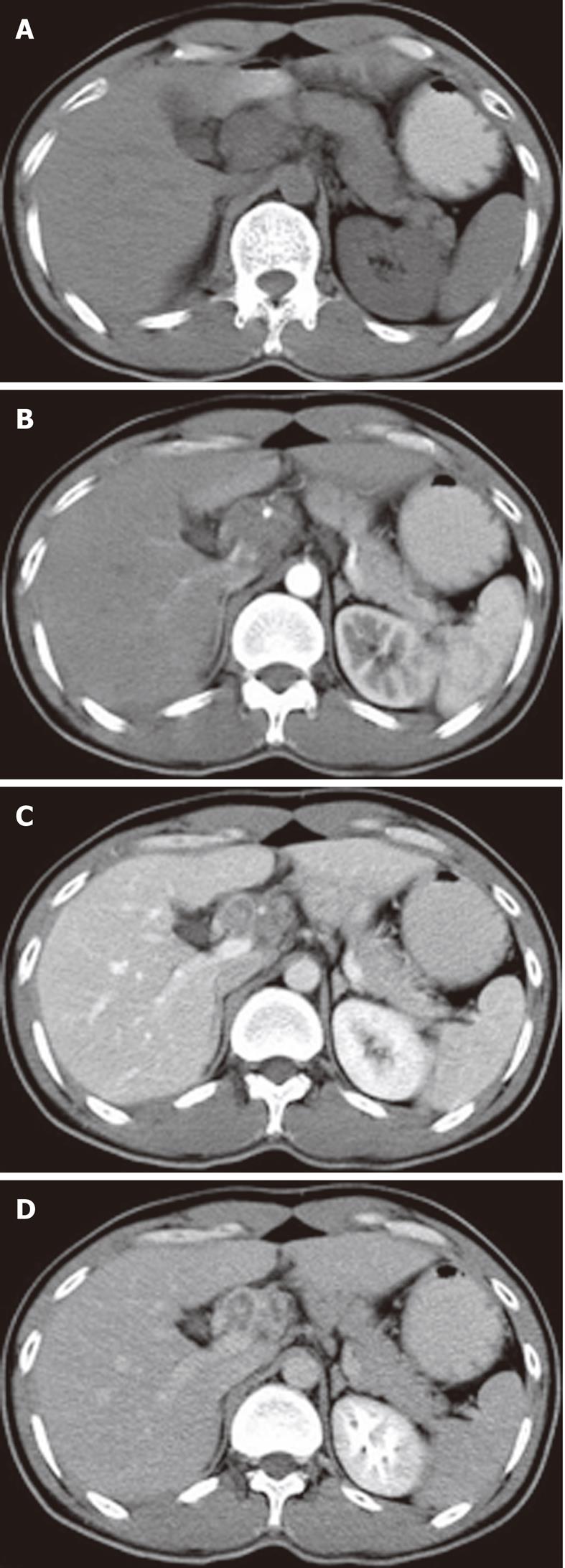
Figure 3 Periportal tuberculous lymphadenopathy.
A: Plain computed tomography scan showing hypodense masses in the hepatic portal region; B-D: After injection of contrast material, continuing peripheral enhancement allows individual lymph nodes to be defined, and the common hepatic artery was embedded within the lesion without definite evidence of stenosis.
- Citation: Zhang M, Li M, Xu GP, Liu HJ. Neoplasm-like abdominal nonhematogenous disseminated tuberculous lymphadenopathy: CT evaluation of 12 cases and literature review. World J Gastroenterol 2011; 17(35): 4038-4043
- URL: https://www.wjgnet.com/1007-9327/full/v17/i35/4038.htm
- DOI: https://dx.doi.org/10.3748/wjg.v17.i35.4038