Copyright
©2011 Baishideng Publishing Group Co.
World J Gastroenterol. Aug 21, 2011; 17(31): 3614-3622
Published online Aug 21, 2011. doi: 10.3748/wjg.v17.i31.3614
Published online Aug 21, 2011. doi: 10.3748/wjg.v17.i31.3614
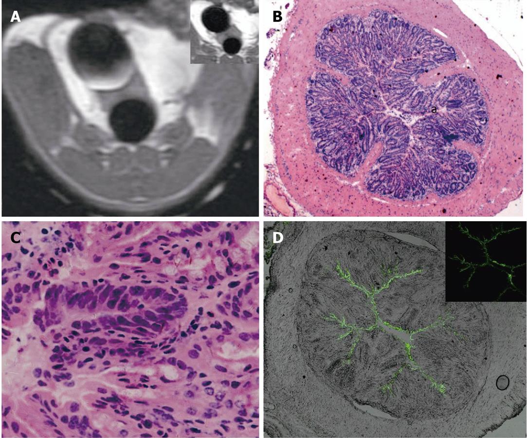
Figure 3 Magnetic resonance and fluorescent images of 1,2-dimethylhydrazine impaired colorectal wall.
A: Axial fluid attenuated inversion recovery (FLAIR) image 5 min after gadopentetate dimeglumine (Gd-DTPA) and fluorescein isothiocyanate (FITC)-loaded solid lipid nanoparticle enema. No mural enhancement was identified. Note the bladder Gd-DTPA deposition (top right: pre-enema FLAIR image ); B, C: Hematoxylin and eosin image of the same slice; nuclear atypia identified; B: × 40; C: × 400. D: Laser-scanning confocal fluorescence microscope image of the post-enema colorectal wall, showing the linear extracellular FITC deposition along the mucosa.
- Citation: Wu T, Zheng WL, Zhang SZ, Sun JH, Yuan H. Bimodal visualization of colorectal uptake of nanoparticles in dimethylhydrazine-treated mice. World J Gastroenterol 2011; 17(31): 3614-3622
- URL: https://www.wjgnet.com/1007-9327/full/v17/i31/3614.htm
- DOI: https://dx.doi.org/10.3748/wjg.v17.i31.3614


