
















©2011 Baishideng Publishing Group Co.
World J Gastroenterol. May 28, 2011; 17(20): 2580-2584
Published online May 28, 2011. doi: 10.3748/wjg.v17.i20.2580
Published online May 28, 2011. doi: 10.3748/wjg.v17.i20.2580
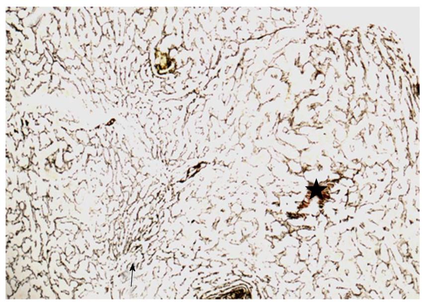
Figure 2 Liver biopsy, reticulin silver impregnation, original magnification, 100 ×.
Hyperplastic parenchymal nodules with thickened liver-cell plates are seen (black star), whereas the parenchymal cells adjacent to the nodules are compressed and atrophic (black arrow). This growth pattern is not accompanied by fibrosis.
- Citation: Gentilucci UV, Gallo P, Perrone G, Vescovo RD, Galati G, Spataro S, Mazzarelli C, Pellicelli A, Afeltra A, Picardi A. Non-cirrhotic portal hypertension with large regenerative nodules: A diagnostic challenge. World J Gastroenterol 2011; 17(20): 2580-2584
- URL: https://www.wjgnet.com/1007-9327/full/v17/i20/2580.htm
- DOI: https://dx.doi.org/10.3748/wjg.v17.i20.2580