
















©2011 Baishideng Publishing Group Co.
World J Gastroenterol. Apr 7, 2011; 17(13): 1666-1673
Published online Apr 7, 2011. doi: 10.3748/wjg.v17.i13.1666
Published online Apr 7, 2011. doi: 10.3748/wjg.v17.i13.1666
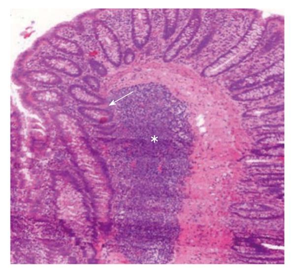
Figure 2 3D reconstruction of a human colonic surgical sample (MIRAX Viewer, 3D, 3DHISTECH Ltd.
, Budapest). A large subepithelial isolated lymphoid follicle (white star) can be seen. Colonic crypts (white arrow) with no connection to the luminal surface “outgrow” from the isolated lymphoid follicle.
- Citation: Sipos F, Műzes G. Isolated lymphoid follicles in colon: Switch points between inflammation and colorectal cancer? World J Gastroenterol 2011; 17(13): 1666-1673
- URL: https://www.wjgnet.com/1007-9327/full/v17/i13/1666.htm
- DOI: https://dx.doi.org/10.3748/wjg.v17.i13.1666