
















©2010 Baishideng Publishing Group Co.
World J Gastroenterol. Oct 7, 2010; 16(37): 4725-4732
Published online Oct 7, 2010. doi: 10.3748/wjg.v16.i37.4725
Published online Oct 7, 2010. doi: 10.3748/wjg.v16.i37.4725
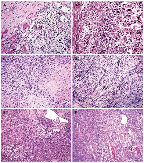
Figure 2 Histology showing residual hepatocytes and bile ducts in the tumor (A), giant cells containing eosinophilic hyaline globules in the cytoplasm (B), loose oedematous myxoid matrix with sparse stellate atypical mesenchymal cells (C), fibroblast-like fascicles (D), angiosarcoma-like cells (E) and pericytoma-like and rhabdomyosarcoma-like cells (F) in compact areas (HE, × 400).
- Citation: Li XW, Gong SJ, Song WH, Zhu JJ, Pan CH, Wu MC, Xu AM. Undifferentiated liver embryonal sarcoma in adults: A report of four cases and literature review. World J Gastroenterol 2010; 16(37): 4725-4732
- URL: https://www.wjgnet.com/1007-9327/full/v16/i37/4725.htm
- DOI: https://dx.doi.org/10.3748/wjg.v16.i37.4725