
















©2010 Baishideng.
World J Gastroenterol. Jan 21, 2010; 16(3): 395-397
Published online Jan 21, 2010. doi: 10.3748/wjg.v16.i3.395
Published online Jan 21, 2010. doi: 10.3748/wjg.v16.i3.395
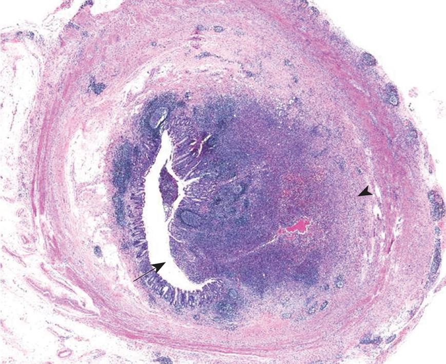
Figure 3 Microscopy of appendiceal actinomycosis.
An abscess composed of chronic and acute inflammatory cells was observed in a mass-like lesion (arrow), from the mucosal surface to the superficial submucosa (arrowhead) (HE, × 10).
- Citation: Lee SY, Kwon HJ, Cho JH, Oh JY, Nam KJ, Lee JH, Yoon SK, Kang MJ, Jeong JS. Actinomycosis of the appendix mimicking appendiceal tumor: A case report. World J Gastroenterol 2010; 16(3): 395-397
- URL: https://www.wjgnet.com/1007-9327/full/v16/i3/395.htm
- DOI: https://dx.doi.org/10.3748/wjg.v16.i3.395