
















©2010 Baishideng.
World J Gastroenterol. Aug 7, 2010; 16(29): 3727-3730
Published online Aug 7, 2010. doi: 10.3748/wjg.v16.i29.3727
Published online Aug 7, 2010. doi: 10.3748/wjg.v16.i29.3727
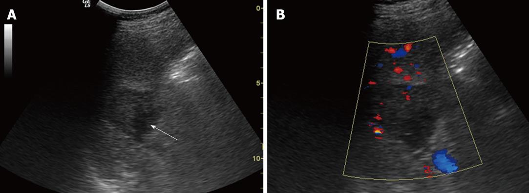
Figure 1 Conventional sonographic findings of sclerosing angiomatoid nodular transformation in a 36-year-old man.
A: B mode ultrasonography demonstrates a hypoechoic lesion (arrow) in the spleen; B: Color Doppler flow imaging shows a low color-flow signal in the lesion.
- Citation: Cao JY, Zhang H, Wang WP. Ultrasonography of sclerosing angiomatoid nodular transformation in the spleen. World J Gastroenterol 2010; 16(29): 3727-3730
- URL: https://www.wjgnet.com/1007-9327/full/v16/i29/3727.htm
- DOI: https://dx.doi.org/10.3748/wjg.v16.i29.3727