















©2010 Baishideng.
World J Gastroenterol. Jun 21, 2010; 16(23): 2851-2866
Published online Jun 21, 2010. doi: 10.3748/wjg.v16.i23.2851
Published online Jun 21, 2010. doi: 10.3748/wjg.v16.i23.2851
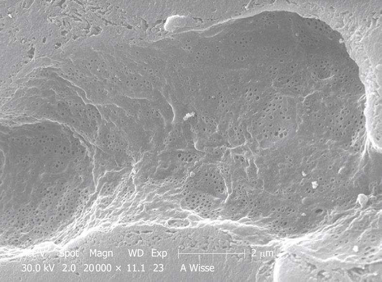
Figure 16 SEM micrograph of another portal-vein-perfused rabbit liver, which shows a complete intact sinusoid, and apparently no gaps have been formed.
Fenestrae occur in sieve plates (groups), the space of Disse seems to be filled by microvilli of the parenchymal cells.
- Citation: Wisse E, Braet F, Duimel H, Vreuls C, Koek G, Olde Damink SW, van den Broek MA, De Geest B, Dejong CH, Tateno C, Frederik P. Fixation methods for electron microscopy of human and other liver. World J Gastroenterol 2010; 16(23): 2851-2866
- URL: https://www.wjgnet.com/1007-9327/full/v16/i23/2851.htm
- DOI: https://dx.doi.org/10.3748/wjg.v16.i23.2851