
















©2010 Baishideng.
World J Gastroenterol. Jun 14, 2010; 16(22): 2735-2742
Published online Jun 14, 2010. doi: 10.3748/wjg.v16.i22.2735
Published online Jun 14, 2010. doi: 10.3748/wjg.v16.i22.2735
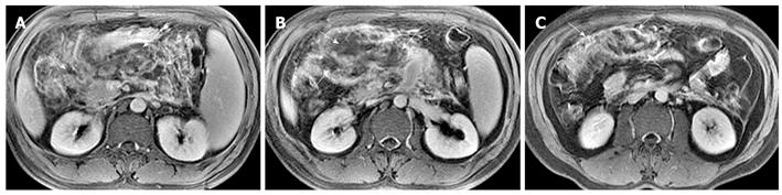
Figure 3 34-year-old man with acute pancreatitis.
Contrast-enhanced axial T1-weighted images show peripancreatic inflammation extension (arrows) along the mesenteric adipose tissue region (A, B). There is addition irregular thickening and enhancement of the adjacent intestinal wall (arrows, C).
- Citation: Xiao B, Zhang XM, Tang W, Zeng NL, Zhai ZH. Magnetic resonance imaging for local complications of acute pancreatitis: A pictorial review. World J Gastroenterol 2010; 16(22): 2735-2742
- URL: https://www.wjgnet.com/1007-9327/full/v16/i22/2735.htm
- DOI: https://dx.doi.org/10.3748/wjg.v16.i22.2735