
















©2009 The WJG Press and Baishideng.
World J Gastroenterol. Mar 7, 2009; 15(9): 1134-1137
Published online Mar 7, 2009. doi: 10.3748/wjg.15.1134
Published online Mar 7, 2009. doi: 10.3748/wjg.15.1134
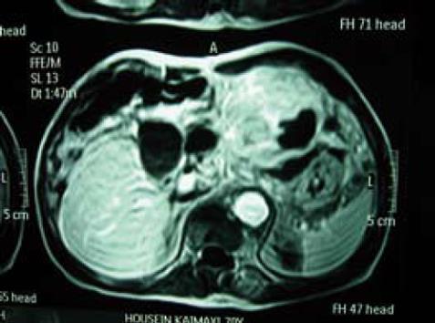
Figure 1 MRI scan demonstrates the presence of a large tumor, 15 cm in diameter, originating from the inferior surface of liver segments II and III.
It is mainly solid with areas of tissue necrosis and shows direct invasion of stomach and pancreas.
-
Citation: Korkolis DP, Aggeli C, Plataniotis GD, Gontikakis E, Zerbinis H, Papantoniou N, Xinopoulos D, Apostolikas N, Vassilopoulos PP. Successful
en bloc resection of primary hepatocellular carcinoma directly invading the stomach and pancreas. World J Gastroenterol 2009; 15(9): 1134-1137 - URL: https://www.wjgnet.com/1007-9327/full/v15/i9/1134.htm
- DOI: https://dx.doi.org/10.3748/wjg.15.1134